|
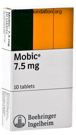
Meloxicam dosages: 15 mg, 7.5 mg
Meloxicam packs: 60 pills, 90 pills, 120 pills, 180 pills, 270 pills, 360 pills, 240 pills
Purchase 7.5 mg meloxicam fast deliveryThe process was initially described with use of iliac crest autogenous bone as an interbody graft arthritis medication for knees order meloxicam online pills. Subsequently arthritis medication voltaren buy meloxicam 15mg amex, nevertheless psoriatic arthritis diet uk purchase meloxicam online pills, allograft bone and different bone graft substitutes have been used with great success rheumatoid arthritis treatment new zealand purchase meloxicam 7.5 mg visa. Although using instrumentation increased fusion charges, it also added the chance of implant-related problems, such as screw and plate breakage and pullout. A high-speed burr, an anterior cervical retraction system, and loupe magnification (preferred) or microscope should be available. A small rolled towel or inflatable intravenous bag is positioned behind the scapula. B, Sagittal picture of a computed tomographic myelogram exhibits the C5-C6 disc herniation (solid white arrow). This gel pad is wrapped across the arm to pad the bony prominences, notably across the elbow and ulnar nerve, before tucking the arm. Extension is achieved with an inflatable intravenous bag or a rolled towel placed throughout the upper back, beneath the scapula. The clavicle and the sternal notch are marked with a marking pen, as is the left-sided transverse incision, on this case, for a C5-C6 method. Lift the platysma with pickups and divide with cautery in line with the skin incision. Scissors or electrocautery adopted by a Kittner dissecting sponge can be utilized to dissect the prevertebral fascia off of the anterior cervical backbone. A needle ought to be placed within the involved disc house, and an intraoperative radiograph or fluoroscopic image should be obtained to affirm the proper degree. The delicate tissues and longus coli muscular tissues could be dissected off of the anterior cervical backbone at the involved level. Discectomy, Fusion, and Instrumentation the anterior annulus is incised with a scalpel. The intervertebral disc and cartilaginous materials is then d�brided with a collection of curved and straight curettes and pituitary rongeurs back to bleeding endplate bone, working from anterior to posterior and out to the uncovertebral joints bilaterally. The white line signifies the situation of the sternocleidomastoid muscle, the black line signifies the location of the clavicle, and the white circle indicates the placement of the sternal notch. As a common guideline, the incision for a C6-C7 approach is made 2 fingerbreadths above the clavicle, the incision for a C5-C6 approach is made 3 fingerbreadths above the clavicle, and so on. As extra anatomical guides: 1, the cricoid cartilage typically sits instantly anterior to the C6 vertebral physique; 2, the carotid tubercle that extends off of the C6 vertebral body can oftentimes be palpated, especially in skinny patients; 3, the thyroid cartilage usually sits instantly anterior to the C4-C5 disc house; and four, the hyoid sometimes sits immediately anterior to the C3 vertebra. The strategy is carried out medial to the sternocleidomastoid muscle (dashed white circle) and the carotid sheath containing the inner jugular vein (dotted white circle) and the carotid artery (solid white circle) and lateral to the trachea (solid red circle) and the esophagus (dashed red circle). The longus coli muscle tissue (dashed white arrow) need to be dissected off of the anterior aspect of the vertebral our bodies (open white arrow). The trachea and esophagus are retracted medially, and the sternocleidomastoid muscle and contents of the carotid sheath are retracted laterally. Umbilical tape (30-inch) can be tied across the retractors bilaterally, and with the untied ends thrown off of the sterile area, weights can be tied to the unsterile end and hung from the table to assist stabilize the retractors during the procedure. The vertebral endplates are prepared again to bleeding bone with a rasp or a highspeed burr. A bone graft (iliac crest autograft or structural allograft) of acceptable dimension is selected and impacted into the intervertebral house with a tamp and a mallet. The plate then is positioned over the vertebral our bodies, and a drill is used to drill screw holes. The screws in the proximal vertebrae should be no much less than 5 mm from the adjoining stage disc area to decrease the risk of developing adjoining stage ossification of the disc. In addition to the preoperative dose of antibiotics given inside 1 hour before skin incision, sufferers should obtain 24 hours of antibiotics after surgery. Sequential compression devices on the bilateral lower extremities and early ambulation are beneficial to minimize the risk of postoperative deep venous thrombosis. The drain is often maintained till postoperative day 1 or until the output is lower than 30 mL per 8-hour shift. Patients are sometimes pretty mobile after this process and can expect to go house on postoperative day 1. Although uncommon, the devopment of a hematoma in the early postoperatve interval could cause airway compromise or neurologic decline. Education of the nursing workers and shut statement is important in detecting these issues early to prevent potentially devastating penalties. The most common points after surgical procedure are related to surgical website pain and swelling, together with sore throat, dysphonia, and dysgphagia, all of which usually steadily enhance over a period of days to weeks. Intravenous or oral steroids could be helpful in managing these symptoms in the early postoperative interval. The addition of lateral flexion and extension radiographs at subsequent follow-up visits might help determine whether or not the bony fusion is solid. Patients can return to their actions of daily dwelling inside a couple of days of surgery. Neck Disability Index, short form-36 bodily component abstract, and pain scales for neck and arm pain: the minimal clinically necessary distinction and substantial medical benefit after cervical backbone fusion. Anterior cervical decompression and arthrodesis for the remedy of cervical spondylotic myelopathy. Radiculopathy and myelopathy at segments adjoining to the location of a previous anterior cervical arthrodesis. Dysphagia and soft-tissue swelling after anterior cervical surgery: a radiographic evaluation. Cervical disc arthroplasty in contrast with arthrodesis for the treatment of myelopathy. Anterolateral cervical disc removal and interbody fusion for cervical disc syndrome. Although most are thought of nondisplaced and could additionally be treated successfully with nonoperative managment, up to 20% require surgical fixation. Proximal humerus fractures follow a bimodal distribution, with the bulk occurring in the aged osteoporotic affected person inhabitants and a 3: 1 predilection toward female patients over males. Patients with proximal humerus fractures present typically with pain and swelling and impaired range of movement in the shoulder after a mechanical fall onto an outstretched arm. Historically, surgical remedy of proximal humerus fractures has been profitable with regard to useful end result but has additionally carried a high price of postoperative issues. Early strategies of fixation with nonlocking plate and screw fixation resulted in hardware failure, lack of fixation, and either nonunion or malunion. Advances in implant design, particularly the development of proximal humerus locking plates, have improved outcomes and decreased complication rates, leading to a current dramatic rise within the fee open discount inside fixation. Absolute indications for operative fixation of proximal humerus fractures include open fractures with or with out neurovascular damage and unstable displaced intraarticular or periarticular fractures that stop early vary of motion and subsequently confer vital useful impairment. When contemplating surgical intervention, a quantity of elements have to be considered, including patient physiologic age, bone high quality, fracture pattern, humeral head vascularity, present useful status, affected person comorbidities, patient expectations, and surgeon experience. Currently, no true gold standard exists for the surgical remedy of proximal humerus fractures. Goals of surgical fixation embrace anatomic reduction with secure fixation to permit for early vary of motion. Ideally, treatment is geared to reduce posttraumatic arthritis and impingement and to reestablish preoperative power and range of motion. The affected person should be positioned as far lateral on the table, toward the operative aspect, as attainable to maximize access to the shoulder. A plastic drape is placed from medial to lateral, just above the nipple line, traversing beneath the axilla. The shoulder then is scrubbed with chlorhexidine and patted dry with a sterile blue towel. The skin is prepared with the cleansing agent of selection (providone-iodine or chlordexidine gluconate+isopropyl alcohol), extending anteromedially to the sternum, inferiorly to the plastic drape, superiorly to simply above the crest of the neckline, and as far posterior as the desk allows. The first is placed beneath the axilla and extends both posteriorly and anteriorly throughout the chest.
Diseases - Dysmorphism abnormal vocalization mental retardation
- Myopathy, myotubular
- Neuraminidase deficiency
- Hypoprothrombinemia
- Nephrolithiasis type 2
- Amelogenesis imperfecta
- Congenital short bowel

Buy meloxicam canadaDuring the primary 15 min after injection of local anesthetics arthritis treatment breakthrough trusted meloxicam 15mg, patients must be rigorously monitored for early identification and remedy of native anesthetic-induced neurotoxicity arthritis relief plus limited buy meloxicam 7.5mg cheap. Furthermore early onset arthritis in neck buy discount meloxicam 7.5 mg line, native anesthetics can have vital adverse chronotropic results arthritis pain glucosamine chondroitin order meloxicam 15mg mastercard. To this finish, a recent paper on the anesthetic administration for awake craniotomy described a case in which the use of native anesthetic infiltration combined with intravenous antihypertensive brokers resulted in important bradycardia and complete atrioventricular coronary heart block [26]. Neurotoxicity and cardiotoxicity must be treated with intravenous infusion of intralipid emulsion (intralipid 20% 1. Another complication of local anesthetic infiltration is transient facial palsy after auriculotemporal nerve block. The precise explanation for the transient postoperative facial nerve palsy after auriculotemporal nerve block is unknown and certain multifactorial. Many hypotheses have been suggested concerning the etiology of transient facial palsy, including direct nerve harm, nerve compression from a hematoma, edema, or native anesthetic injection that will lead to neural ischemia and harm, and direct neurotoxic results of native anesthetics [26�28]. Providing too much sedation may find yourself in an uncooperative patient with or with out respiratory despair, whereas providing too little sedation ends in an uncomfortable and agitated patient, which might find yourself in arterial hypertension and tachycardia. Providing sufficient sedation could make awake anesthesia for craniotomy less physically and emotionally annoying than common anesthesia [20, 29]. Intravenous anesthetic brokers that have been described for sedation protocols include propofol�fentanyl, propofol�remifentanil, and dexmedetomidine (Table 12. A bispectral index monitor can be helpful when titrating a propofol infusion to a goal conscious state [18, 20]. In patients present process awake craniotomy, the anesthetic protocol should also obtain enough postoperative ache control. Remifentanil is a well-tolerated opioid that gives good intraoperative pain control. However, it ought to be famous that discontinuation of remifentanil has been associated with a quantity of postoperative issues, including hyperalgesia, hypertension, and tachycardia [20, 31, 32]. Cortical resection tailored to awake, intraoperative ictal recordings and motor mapping within the remedy of intractable epilepsia partialis continua: technical case report. Nonlesional central lobule seizures: use of awake cortical mapping and subdural grid monitoring for resection of seizure focus. Effects of sevoflurane and isoflurane on electrocorticographic actions in patients with temporal lobe epilepsy. Discrepancies between preoperative stereoencephalography language stimulation mapping and intraoperative awake mapping during resection of focal cortical dysplasia in eloquent areas. Awake surgical procedure with steady motor testing for resection of brain tumors within the primary motor area. Anesthesia management of awake craniotomy carried out underneath asleep-awake-asleep method utilizing laryngeal masks airway: report of two instances. Maintenance of normotension or slight hypotension is important to reduce bleeding and brain swelling that can happen during brain exposure, and to obtain surgical hemostasis. Severe arterial hypertension and tachycardia are associated with a major risk of postoperative intracranial hemorrhage and myocardial ischemia, respectively, and ought to be prevented or promptly handled [20]. Several agents can be utilized to decrease blood pressure, including beta blockers (esmolol, metoprolol, atenolol), calcium-channel blockers (diltiazem and verapamil), alpha blockers (urapidil) combined alpha and beta blockers (labetalol), and alpha-adrenergic receptor agonists (clonidine). Conclusion Awake craniotomy is a well-tolerated procedure that requires an intensive data of underlying neuroanesthesia principles, as well as specific methods of native anesthetic scalp blockade, advanced airway management, a dedicated sedation�analgesia protocol, and skillful management of systemic and cerebral hemodynamics. Recent trends within the anesthetic management of craniotomy for supratentorial tumor resection. Awake insertion of the laryngeal masks airway using topical lidocaine and intravenous remifentanil. The impact of scalp block and native infiltration on the haemodynamic and stress response to skull-pin placement for craniotomy. Combination of bupivacaine scalp circuit infiltration with common anesthesia to control the hemodynamic response in craniotomy patients. Effect of a subanesthetic dose of intravenous ketamine and/or local anesthetic infiltration on hemodynamic responses to skull-pin placement: a prospective, placebo-controlled, randomized, double-blind research. Prevention and treatment of local anesthetics-induced complete atrioventricularblock throughout awake craniotomy. Transient facial nerve palsy after auriculotemporal nerve block in awake craniotomy sufferers. Training anesthesiology residents in providing anesthesia for awake craniotomy: learning curves and estimate of needed case load. Rapid improvement of tolerance to analgesia throughout remifentanil infusion in people. Management of hypertensive emergencies in acute mind disease: evaluation of the remedy results of intravenous nicardipine on cerebral oxygenation. To higher achieve it, the modern anesthesiologist should use medication with a predictable and favorable profile: a fast onset and offset, secure and fast induction, early restoration, easily titratable and with out - or minimal - antagonistic or unwanted effects. Pending the event of latest and "almostideal" medicine, the emergence and enchancment of techniques and units that permit their protected and accurate supply, have contributed to the development of our practice. Thus, the debate continues on intravenous and inhalation anesthesia concerning well-defined clinical endpoints. These are mainly associated to the velocity and recovery of anesthesia, hemodynamic adjustments, operative circumstances, postoperative nausea and vomiting, restoration of psychomotor and cognitive perform, and discharge from hospital. Costa (*) Department of Anesthesiology, Centro Hospitalar Lisboa Central, Lisbon, Portugal e-mail: martinsdacosta. Lobo Department of Anesthesiology, Hospital Geral de Santo Ant�nio � Centro Hospitalar do Porto, Porto, Portugal � Springer International Publishing Switzerland 2017 Z. Furthermore, research on long-term outcomes has shown that intravenous anesthesia with propofol could additionally be more favorable concerning cancer recurrence after surgical procedure [3]. Although several contradictory outcomes have been printed regarding immunosuppression, plainly propofol attenuates surgical stress response without impairment of pure killer cell activity and resulting in less vital immunosuppression [4]. Aware of the advantages described, financial influence has been a topic of nice curiosity with inadequate research till now. Therefore, all costs and benefits should be thought-about, related not solely to the approach but additionally the establishment, the patient, and the society [5]. Nevertheless, we ought to always not forget the safety and straightforward titration of risky anesthetics and acknowledge the deserves of each techniques [6]. Clear indications for the use of each method are missing and the choice seems to be extra associated to the individual expertise and familiarity with the method than based mostly on revealed studies, without forgetting the provision of the tools. The apply of target-controlled anesthesia is predicated on elementary pharmacokinetic and pharmacodynamic ideas which deserve a detailed evaluation. Drug elimination by metabolism from the central compartment corresponds to the constant K10. They are the- oretical volumes that can be remotely thought because the blood volume (V1), a "vessel rich" compartment (V2) and a "vessel poor" compartment (V3). In every model, the dimensions of the volumes of distribution reflects the solubility of the drug in that particular compartment. For pharmacokinetic/pharmacodynamic modeling, an effect-site compartment could be added. Due to its negligible volume, the rate constants for motion out and in of this compartment are the same (K1e = Keo). In the previous many years, pharmacokinetic models have been developed and improved, allowing better understanding of the habits of various drugs and making possible the adjustment to the individual kinetics. It is outlined as the concentration at regular state multiplied by the clearance worth (Cl). Generally, it might be calculated knowing the dose current within the physique (D) and its plasma focus (C): Vd = D / C (13. Half Life (t1/2) is the time taken for the concentration of a drug to drop by half its value. However, since most drugs are distributed in more than one compartment and clearance from each occurs at totally different rates, the drug could have more than one t1/2. Decremental time is an identical concept that defines the time required for blood focus to decrease to a desired proportion. Both ideas are helpful predictors of drug focus decline after an infusion is stopped. If another bolus is required to enhance the goal focus, New Loading Dose = (Cf - Ci) � Vd; (13. Clearance (Cl) describes the amount in which the drug is eliminated during a unit of time (mL/ min or mL/hr). For this reason it turns into obvious that the primary effect is delayed in addition to the drug focus at the effect web site when compared to the plasma peak focus.
Buy meloxicam online nowThe assembled syringe and needle then are placed into the stress monitor arthritis hot feet order meloxicam 15 mg visa, and the duvet is closed and locked arthritis dx code discount 15 mg meloxicam visa. A large C-arm is positioned on the contralateral side of the affected extremity within the setting of deliberate fracture fixation arthritis zapper purchase meloxicam 7.5mg mastercard. A small bump could additionally be positioned under the ipsilateral hip to faciliate access to the operative extremity arthritis in joints of fingers generic meloxicam 7.5 mg line. A0-Compartment syndrome, unspecified Prepping and Draping the affected extremity is prepped and drapped in a sterile manner. Compartment Pressure Measurement Use a marking pen to mark relevant anatomic landmarks, together with the define of the fibula, the fibular head, and the lateral malleolus. Superficial posterior compartment: Pressure measurement is taken over the posteromedial aspect of the gastrocsoleus. Deep posterior compartment: Pressure measurement is taken on the posteromedial border of the tibia in the distal one half of the decrease leg. The anterolateral incision is made between the fibula and the anteiror tibial crest, simply anterior to the intermuscular septum between the anterior and lateral fascial compartments. Double-Incision Technique the double-incision approach is probably the most commonly used approach due to its relative technical ease, predictable compartment launch, and safety. A longitudinal incision is placed midway between the fibula and the anterior tibial crest. Incision length is roughly 5 cm distal from the fibular head to 5 cm proximal to the lateral malleolus. A small transverse incision must be made within the fascia to permit for direct visualization of the intermuscular septum between the anterior and lateral compartments. The tips of the scissors then could be inserted by way of the previously established lease within the fascia. Separate longitudinal incisions should be made in each the anterior and the lateral compartments to keep away from iatrogenic harm to the intermuscular septum and to the superficial peroneal nerve. Transverse incisions are revamped the fascia of the anterior and lateral compartments, under which the intermuscular septum is recognized. Care must be taken to not harm the superficial peroneal nerve, which lies simply posterior to the septum and could additionally be encountered approximately 10 cm proximal to the lateral malleolus, the place it programs from lateral to anterior compartments. The superficial and deep posterior compartments are accessed by way of a longitudinal incision made on the posteromedial facet of the decrease leg, approximately 2. Identification of the saphenous vein and nerve is carried out, crossing the wound from posterior to anterior. Once recognized, anterior retraction of the neurovascular buildings is performed. Decompression of the complete superficial posterior compartment is carried out by releasing the fascia overlying the whole gastrocsoleus complex. Decompression of the entire deep posterior compartment is carried out by releasing the fascia overlying the flexor digitorum longus. Dissection by way of subcutaneous tissue is performed, as is identification and incision of the fascia to expose the intermuscular septum. Similar to the method for the anterolateral incision, a small transverse incision should be made within the fascia, which permits for direct visualization of the intermuscular septum between the deep and superficial compartments. After release of the deep compartment fascia, the scissors ought to be oriented proximally by way of, and deep, to the soleus bridge to enable for launch of the soleus attachment to the tibia. Full fascial release of the anterior and lateral compartments is carried out as described beforehand. The skin is undermined posteriorly, and the musculature of the lateral compartment is carefully elevated and retracted to permit for identification of the posterior intermuscular septum. The interval between the lateral and the superficial posterior compartments is recognized and developed by proximally detaching the soleus from the fibula and subperiosteally dissecting the flexor hallicus longus from the fibula. Tissue is retracted posteriorly, and the fascial attachment of the tibialis posterior is recognized and incised longitudinally. D�bridement and Closure Regardless of the discharge technique used, a devoted d�bridement with elimination of as a lot necrotic tissue, if current, as possible is imperitive. Many choices exist for closure, together with delayed major closure, cut up thickness skin grafting, dermotraction, and vacuum-assisted wound closure. Postoperative monitoring for systemic signs of compartment syndrome is established, and supportive postoperative care is run as is clinically indicated. Often, patients are brought again to the working room 2 to 3 days after the index procedure for repeated d�bridements or delayed closure. This determination is predicated on the state of the incisions, the general condition of the affected extremity, and the postoperative clinical status of the patient. After this appointment, sufferers return to the office for orthopaedic evaluation at 6 weeks, three months, 6 months, and 1 yr status after index procedure. If postoperative issues occur, this schedule could also be adjusted accordingly such that acceptable care is provided. Dermotraction: an efficient technique for the closure of fasciotomy wounds: a preliminary report of fifteen patients. Wound closure of leg fasciotomy: comparison of vacuumassisted closure vs shoelace method: a randomised examine. The pathophysiology of the anterior tibial compartment syndrome: an experimental investigation. Comparison of fasciotomy wound closures utilizing traditional dressing changes and the vaccum assisted closure device. Any articular fracture of the proximal tibia is assessed as a tibial plateau fracture. This broad classification allows for a variety of severity of injury to the articular floor, fracture extension to the tibial metaphysis and diaphysis, and gentle tissue injury. An orthopaedic surgeon who manages these accidents should have an understanding of how these harm traits affect operative management. High-energy tibial plateau fractures usually happen in young, active individuals with robust bone stock, and low-energy accidents, such as a fall from standing, typically happen in patients with osteoporosis. The magnitude and path of the pressure to the extremity affects the damage pattern. Schatzker in 1974 described a classification for articular fractures of the proximal tibia that led to the terms surgeons use to describe these accidents. Learning the language to describe these fractures is essential for communication between surgeons. The common phrases used to describe this fracture pattern are split, despair, or split-depression, along with the placement of the fracture, together with medial condyle, lateral condyle, or bicondylar. Split depression of the lateral condyle is the most typical fracture pattern due to the 5-degree to 7-degree regular alignment of the knee and the propensity of people to be struck on the lateral side of the leg. In this pattern, the medial collateral ligament acts as a hinge for the valgus second at the knee, resulting in failure of the lateral articular floor. Severe gentle tissue accidents are sometimes associated with bicondylar fractures, fracturedislocations, and metaphyseal-diaphyseal dissociated patterns. These sufferers often need spanning exterior fixation to permit for gentle tissue therapeutic earlier than open discount and inside fixation. Open wounds and fracture blisters are indications for spanning exterior fixation with delayed inner fixation or definitive external fixation in choose circumstances. Compartment syndrome generally occurs in sufferers with high-energy fracture patterns. Stark and colleagues reported compartment syndrome after placement of spanning external fixation in 18% of bicondylar tibial plateau fractures and in 53% of medial plateau fracture-dislocations. This highlights the importance of continued monitoring for compartment syndrome in patients with high-energy patterns, particularly fracture-dislocations. Open discount and internal fixation of tibial plateau fractures have been reliably carried out with modern strategies since the Nineteen Eighties. F, Bicondylar fracture with proximal tibia fracture dissociating the articular block from the diaphysis. Restoring alignment of the lower extremity has been shown to correlate with improved affected person outcomes. In contrast, the articular floor of the proximal tibia has tolerance of modest deformity.
Buy meloxicam 15mg cheapIn Italy arthritis diet eating plan order meloxicam 15mg online, nevertheless arthritis in fingers numbness generic 7.5 mg meloxicam with amex, fashionable obstetrics has restricted its use and few apply it for concern of problems and medicolegal repercussions arthritis in border collie dogs order meloxicam 15 mg visa. The mother obviously should not have extreme belly subcutaneous panniculus adiposus [1 arthritis simple definition order meloxicam 15 mg on-line,7]. Relative maternal contraindications are gestational age under 36 weeks, maternal pathologies (such as hypertension, diabetes, gestosis, hyperthyroidism, etc. Absolute fetal contraindications as an alternative are the following: twin being pregnant or serious fetal anomalies (hydrocephalus, anencephaly, coronary heart illness, etc. The relative contraindications are the next: delay in intrauterine growth or estimated fetal weight over 4000 grams and incomplete breech variant. Absolute contraindications of fetal adnexa are the next: abnormal placental insertion (placenta praevia), important oligohydramnios (under 50), or untimely rupture of the membranes. To perform this technique the pregnant woman must have an empty bladder, be lying supine with barely flexed legs, and have signed an knowledgeable consent. Some obstetricians prefer the woman to be in a slight Trendelenburg position, and others favor the lateral decubitus place. While the operator lifts the uterine breach together with his or her left hand to increase the out there house, the best hand is used to grab the feet and extract them from the breach, thus extracting the legs. First section of the Mauriceau maneuver: the surgeon, along with his or her proper hand, locates the buccal cavity of the fetus and inserts his or her index and center fingers. The assistant maintains a relentless suprapubic pressure to facilitate the maneuvers of the operator. Initially, the proper upper limb had risen alongside the top; the forearm then dropped behind the occiput toward the higher part of the fetal dorsum. The obstetrician places his or her practically flat arms on the two fetal poles and applies a constant pressure within the try to turn the fetus with an anterior (craniocaudal) or posterior (caudocranial) motion. Some obstetricians prefer to exert pressure solely on the fetal dorsum or only on one fetal pole (breech or cephalic extremity). For this reason the rotation ought to be carried out in utterly protected conditions, with a team ready to intervene should a cesarean delivery turn into essential [19]. After the maneuver, it is recommended to use tocolytics, similar to nifedipine, terbutaline, or atosiban, to chill out the uterus [17�19]. It is therefore recommended to by no means exert too much force during the model [22]. Initially, the upper limb was lowered alongside the facet of the fetus; the forearm then moved behind the dorsum and was pressured to move in an upward course. In some countries, physicians resort to external cephalic version, assisted by tocolytic betamimetics [28]. In trendy obstetrics, breech presentation, generally, results in delivery by cesarean delivery [28,29]. Due to medical and authorized risks tied to vaginal start issues [30], most obstetricians choose abdominal delivery to assisted vaginal supply [31�33]. Generally, cesarean supply of a breech presentation can be planned after the thirty eighth week of gestation and outdoors of labor [34,35]. Maneuvers for supply of the fetus in breech presentation, throughout a cesarean delivery, are the same as those during assisted vaginal delivery. Similarly, external cephalic model maneuvers are principally the identical as those who were performed up to now [8�11] to keep away from fetal damage [40]. It favors the rotation of the fetus from breech to cephalic current and is without specific dangers. Furthermore, model reduces the larger frequency of funicle complications related to breech delivery, by which the incidence of prolapsed funicle is 3�20 occasions that of cephalic delivery [1,7,20]. It has been established that version maneuvers modify the guts fee in 20%�40% of instances. These changes, nonetheless, are transitory and disappear quarter-hour after the maneuvers finish [23]. For this reason at the finish of the procedure anti-D immunoglobulins are administered to women with Rh-negative blood sort. The share of positive outcome of the maneuver, when correctly performed, is round 70%�90% [1,7,24]. The scientific literature stories that after model happens at the thirty seventh week or later, only a few fetuses return to the breech presentation [25]. Planned caesarean supply versus deliberate vaginal start for breech presentation at time period: A randomised multicentre trial. Randomized control trial of external cephalic version with tocolysis in late pregnancy. Nifedipine as a uterine relaxant for external cephalic model: A randomized managed trial. Oral nifepidine versus subcutaneous terbutaline tocolysis for external cephalic version: A double-blind randomised trial. External cephalic model among women with a previous cesarean supply: Report on 36 cases and evaluate of the literature. Safety and efficacy of external cephalic version for ladies with a previous cesarean delivery. Massive fetomaternal hemorrhage following failed external cephalic model: Case report. Interventions for helping to flip term breech babies to head first presentation when utilizing exterior cephalic version. The costs of planned cesarean versus deliberate vaginal start in the Term Breech Trial. Systematic evaluate of opposed outcomes of exterior cephalic model and persisting breech presentation at time period. Planned vaginal delivery versus elective caesarean supply in singleton time period breech presentation: A research of 1116 circumstances. Planned caesarean supply decreases the chance of adverse perinatal outcome because of both labour and supply problems within the Term Breech Trial. Maternal mortality and severe morbidity associated with lowrisk deliberate cesarean supply versus deliberate vaginal delivery at term. Pregnancy outcome after profitable external cephalic model for breech presentation at time period. Vaginal versus cesarean delivery for breech presentation in California: A population-based study. Caesarean supply and proper femur fracture: A uncommon however possible complication for breech presentation. In addition, subjective strain could be applied on the scalp, and when essential, the device may be deactivated manually. In utilizing forceps during a cesarean delivery, larger attention have to be paid to application time and traction methods. The latter, however, have phases which are comparable to these of vaginal delivery. The use of forceps or vacuum extractor should be considered solely when manual maneuvers fail [2�4]. Using forceps Short forceps with crossed branches and sliding mechanism (Pajot or Smellie) or with divergent branches (Suzor) can be utilized. As another, a single forceps branch may be used as a lever, with the pubic symphysis functioning as a fulcrum. There are two various sorts of cephalic presentation, relying on whether the fetal occiput is anterior or posterior. If a caesarean supply is performed to shield the mind of the child from the trauma which would occur vaginally, or for obstetric reasons, particular attention should be paid when eradicating the body and the head. An extraction with devices is always preferable to a widening of the hysterotomy breach because the pedunculi of the uterine arteries may be damaged [1]. Besides these particular instances during a cesarean delivery during which the fetal head needs to be extracted with have an upward concavity in case of anterior occiput and downward concavity in case of posterior occiput. A single department of the forceps may be used when extraction of the pinnacle from the uterine breach proves to be particularly difficult.
Lobelia. Meloxicam. - How does Lobelia work?
- Use by mouth for asthma, bronchitis, cough, and other conditions.Use on the skin for muscle soreness, bruises, sprains, insect bites, poison ivy, ringworm, and other conditions.
- Smoking cessation.
- Dosing considerations for Lobelia.
- Are there any interactions with medications?
- What is Lobelia?
Source: http://www.rxlist.com/script/main/art.asp?articlekey=96260
Order meloxicam 15mg on lineIt was an overall optimistic expertise arthritis facts order genuine meloxicam on line, particularly for some patients who could objectively assess their own labor arthritis support groups purchase meloxicam 15 mg on line. Generation of continuous and accurate knowledge on two essential parameters for the evaluation of development of labor: cervical dilatation and level of the fetal head arthritis pain worse in the morning buy cheap meloxicam 15mg on line. Subjective and inaccurate evaluation of cervical dilatation rheumatoid arthritis complications purchase 15 mg meloxicam amex, as decided by vaginal examinations at intervals of 3�4 hours. Reduction in the number of vaginal examinations, which is most likely not effective or might even be harmful for varied causes. Generation of objective information and definite parameters on the progression of labor, which can be displayed on a pc in the room of the obstetrician-gynecologist on duty, thereby eliminating the necessity for a quantity of telephone calls between medical and nursing employees. Ability of the staff to make timely calls in case of imminent start (obstetrician�gynecologist, obstetrician� anesthetist, obstetrician�neonatologist). Up to now in more than 300 cases analyzed, the risk of an infection and bleeding is only theoretical, while the risk of cervical abrasion is very small (<0. An extra monitor would be used throughout labor, making delivery even less natural and more medicalized. The benefits of this technique, though, clearly outweigh these theoretical obstacles. Possible future applications of cervicometry the current applications of this method are considerable, but in the close to and imminent future these devices may considerably change how labor is carried out. In a preliminary research on the physiology of individual contractions [53], the Barnev cervicometer was used to evaluate the effect of individual contractions on dilation modifications and the position of the presenting part. These preliminary knowledge indicate a big shift in the perspective by obstetricians concerning cervical dilations and the place of the top in response to uterine contractions. The information also suggest that this model could be very helpful in figuring out the exact second by which the lively phase begins and dilation is complete. Early detection of contractions not useful to dilation can be added to the cartographic parameters and could be concurrent with the analysis of gradual dilation or poor descent of the fetal head. These data may lead to early obstetric interventions, thereby probably reducing the want to resort to a cesarean delivery. In gentle of the above, the authors recommend that evaluation of speedy adjustments in cervical dilatation and/or engagement of the presenting half with a cervicometer might be a superb diagnostic and therapeutic foundation. As talked about, oxytocin, beta-agonist with a really short half-life, could also be used based on the frequency and length of contractions, each surrogate parameters within the evolution of childbirth labor. Cervical dilatation and descent of the pinnacle are, nonetheless, the 2 most dependable parameters for monitoring the development of labor. It follows from the above that both may be evaluated with computerized cervicometry. The authors of this chapter believe that, in a not-toodistant future, the dosage of oxytocin may be decided from short-term modifications in cervical dilation and within the References 233 position of the top (both indicated by the cervicometer). This would eliminate the current long vaginal examination interval of 2�4 hours, in addition to decreasing the variety of vaginal examinations and resulting endometritis and chorioamnionitis. A preliminary investigation of dystocic labor on an animal mannequin has led to the conclusion that this instrument can doubtlessly result in an effective and timely obstetric intervention. Short-term modifications in dilation and in the position of the presenting half, along with the fashions offered by the instrument during the contractions, can present good suggestions for guiding the administration of oxytocin. As a result of its software there would be a discount in vaginal visits, a lower rate of an infection, a reduction within the price of cesarean deliveries, and, most likely, cesarean deliveries carried out early, with all the mandatory indications [50�54]. Dystocia represents about 50% of the causes of operative deliveries and, in particular, of cesarean deliveries, while fetal distress represents 1%�2% of operative deliveries. An enhance in authorized disputes necessitates an objective evaluation of maternal and fetal pathologies, and thus additionally of dystocia. Several studies have confirmed the diagnostic unreliability of vaginal examination, both within the first in addition to in the second stage of labor. Therefore, the need in obstetrics for goal feedback has turn out to be increasingly evident. The use of intrapartum ultrasound has been proposed to make up for the diagnostic inadequacy of the traditional obstetric examination, and this has considerably reduced the error within the prognosis of fetal head place by 40%�70%. Nevertheless, a system that can objectively evaluate the different maternal and fetal delivery variables has not yet been validated. Different instruments, named cervicometers, have been experimented with prior to now, in the try and overcome this downside, but with out significant clinical success. Researchers have achieved important results by using a ultrasound tool and putting transmitters on the abdomen of the patient, so as to produce waves that might be interpreted by special mathematical algorithms in a computer. The improvement of this instrument and the target evaluation of different parameters (currently not assessable) could represent a scientific application system that objectively determines dystocic birth, in the identical method because the cardiotocograph interprets fetal misery. Clinical Benefit Is Not a Factory Determining Acceptability of Screening for Gestational Diabetes. Trends in fetal development amongst singleton gestations within the United States and Canada, 1985 via 1998. Incidence and significance of the unengaged fetal head in nulliparas in early labor. Planned caesarean section versus deliberate vaginal birth for breech 234 Dystocia and cesarean delivery 18. Trends and issues in labor induction in the United States: Implications for clinical follow [see comment]. Birth simulator: Reliability of transvaginal assessment of fetal head station as outlined by the American College of Obstetricians and Gynecologists classification. Comparison of transvaginal digital examination with intrapartum sonography to determine fetal head place earlier than instrumental supply. Accuracy and inter-observer variability of simulated cervical dilatation measurements. International multicenter time period prelabor rupture of membranes research: Evaluation of predictors of clinical chorioamnionitis and postpartum fever in patients with prelabour rupture of membranes at time period. Inaccuracy in cervical dilatation evaluation and the progress of labour monitoring. Measurement of the forces and strains of labour and the motion of sure oxytocic drugs. Electronic cervimeter: A analysis instrument for the study of cervical dilatation in labor. A cervimeter for continuous measurement of cervical dilatation in labour: Preliminary outcomes. Continuous monitoring of cervical dilatation throughout labor by ultrasonic transit time measurements. An ultrasonic gadget for steady measurement of cervical dilatation throughout labor. For this function we begin our evaluation by taking a step back and looking at comparative anthropology. The ratio between fetal head and delivery canal in all anthropoid apes is such that the top of the fetus can progress into the birth canal once cervical dilatation happens, without encountering any of the dimensional limits of the birth canal itself. The human species as an alternative is the only one in which the pinnacle can transfer via the birth canal solely via a complex inner rotation motion. Such a limit, developed during the millions of years which have led to the upright place, has not allowed the mind weight to change (1. We could add to this paradigm another one: fetuses with greater percentiles of somatic growth for our species encounter a second anthropomorphic limitation at childbirth. The second aspect that emerges from comparative anthropology is that delivery in all anthropoid apes is a "non-public" occasion. The feminine when giving delivery distances herself from the group, a lot so that even right now there are very few photographs of childbirth in nature. The selective benefit in hominids offering help to their females at childbirth will increase the variety of youngsters within the group. This has likely resulted in childbirth being, in all human cultures, a social event by which ladies are assisted by third parties, both psychologically and bodily. Even today we can perceive the dramatic impact of childbirth in the human species. In rural areas of Ghana the proportion of rectovaginal fistula for outcomes of arrest within the development at mid-strait is 2% [4,5]. In 2000 the maternal mortality rate was 830 deaths per a hundred,000 girls in Africa, in Asia 330 deaths/100,000 women (not together with Japan and Korea), 240 deaths in Oceania (not together with Australia and New Zealand), in Latin America one hundred ninety deaths, and in the industrialized nations 20 deaths per a hundred,000. The highest variety of deaths in a single yr was in India, with 136,000 expectant moms who died.
Order meloxicam 15 mgBrain Death: Understanding the Process of Brain Death Declaration Through Real-Life Case Scenarios Abhijit Lele and Michael Souter 4 four arthritis pain in dogs order meloxicam cheap. In some sense arthritis in fingers foods to avoid buy meloxicam 7.5 mg with amex, all demise could be thought-about as brain dying in that the sustained cessation of cardiovascular exercise will ultimately give rise to irreversible cessation of brain function king bio arthritis joint relief purchase meloxicam canada. This idea of a dual etiology of death has subsequently unfold throughout most of the world arthritis pain when it rains purchase meloxicam from india. However, an accurate and complete understanding of the diagnosis of demise from neurological causes remains a persistent challenge for medical practitioners in all places. This guide chapter is written to information that understanding, which in turn demands consideration of principles of brain demise declaration within the particular contexts of hypothermia, household refusal to accept the declaration based on spiritual grounds, applicable use of ancillary testing, and understanding post-declaration events. The ideas of the process of declaring mind demise might be discussed within the context of case scenarios the place on analysis of a medical downside, the readers shall be offered with our rationale for management, given the current proof. In essence, this chapter is intended to present the reader with a practical strategy to the declaration of brain demise. It is estimated that mind demise declaration occurs in roughly 5% of sufferers with acute brain injury [9]. Leading causes of mind damage progressing to brain dying include traumatic mind damage, intracranial hemorrhage, and hypoxic/ anoxic-ischemic encephalopathy [10, 11]. Souter 1971 1971 1971 1981 1981 1987 1991 1996 mind dead (Canada, Sweden, Columbia, Chile, Mexico, Panama, and the Russian Federation) [10]. Recently, simulation-based courses have shown to enhance academic efficiency and consequently scale back opportunity for error [13�16]. At the core of declaration is the presence of coma, supported by an sufficient historical past of complaint with radiological findings. Neurological examinations are confounded by hypoxia, hypotension, irregular biochemistry, and affordable suspicion for toxins (either iatrogenic or accidental) (Tables 4. We will present how these criteria can be included in diagnostic determination making, by way of consideration of the following examples. In a current survey of fifty neurological institutions throughout the United States, 42% of all declarations have been carried out by neurologists/neurosurgeons, whereas a majority (65%) of those declarations were performed by resident physicians versus attendings [12]. Around the world, most international locations require a minimal of two physicians to declare patient brain death (ostensibly to avoid error), whereas solely in a couple of can a single doctor pronounce patient An 18-year-old male is concerned in a highspeed motorized vehicle accident, and suffers extreme traumatic brain damage, with extreme facial injuries. He is intubated within the area and is placed in a cervical collar, but without cervical spine fracture on x-ray. Initial neurological examination reveals no cough, gag, or response to noxious stimuli. He has bilateral eyelid edema (rendering pupils troublesome to examine), and blood popping out of his ears. Comprehensive medical evaluation and neurological evaluation type the idea of any brain demise examination [7]. Patients should lack all evidence of responsiveness, with absent eye opening or eye movement to noxious stimuli. There must be absence of brainstem reflexes, corresponding to absence of pupillary response to a bright light documented in both eyes. Usually the pupils are fastened in midsize or dilated (4�9 mm), 4 Brain Death: Understanding the Process of Brain Death Declaration Through Real-Life Case Scenarios fifty seven Table 4. No eye movements should be seen in the 60 s following completion of the irrigation. Oculocephalic reflex (test only when no fractures or instability of cervical spine is apparent) Briskly rotate head 90� lateral from midline (horizontal) and briskly flexion (vertical) head. Cough reflex Stimulate tracheobronchial tree by passing cannula or irrigating endotracheal tube. Hemodynamically steady without cardiac arrhythmias (Systolic blood strain >100 mm Hg either with or without vasopressors). The apnea take a look at must be completed as part of the primary examination during which no other brain operate is demonstrated. The apnea take a look at must be accomplished after the motor response and brainstem reflex testing. Results of trial ought to be documented in medical record, together with length of apneic interval, blood fuel results, and fee and measurable quantity of breaths, if any occurred. Souter Suggested time to mind demise exam Pentobarbital degree 10mcg/ml Phenobarbital stage 10mcg/ml 10�20 h Midazolam 1�12. Also, oculovestibular reflex pre-testing requires demonstration of patency of the external auditory canal, which may be troublesome in our case. Periorbital edema could confound evaluation of eye actions in addition to pupillary reflex. In addition, documentation of absence of corneal reflex, absence of facial muscle actions to noxious stimuli at level of temporomandibular joints or supraorbital and supratrochlear ridges, absence of pharyngeal (gag) reflex and absence of tracheal (cough) reflex are all required. For complete description of testing for coma and brainstem reflexes, refer to Table 4. A 43-year-old male sustains motorized vehicle accident with severe traumatic mind injury, and pulmonary contusions. Consideration of the pre-apnea arterial blood pH might help distinguish between these causes. A lower pH would indicate respiratory acidosis, and air flow must be adjusted to first produce normocapnia and the test then performed. Thus, close consideration must be paid to P/F ratio and systolic blood stress prior to the initiation of apnea check. Consequently, on this clinical scenario, his hypotension would confound neurologic evaluation, while PaO2/FiO2 ratio beneath 200 would rule out secure efficiency of an apnea take a look at. A 38-year-old feminine is present process apnea testing in the course of the means of mind death testing. The respiratory therapist places the patient on a T-piece circuit with reservoir bag, and the patient undergoes 6 min of apnea. A 58-year-old gentleman underwent induced hypothermia treatment to 33 �C for treatment of hypoxic neurological injury following a cardiac arrest. Clinical examination demonstrated the absence of eye opening, verbal response, and motor response, in addition to absence of all mind stem reflexes, with apnea on the ventilator. Twelve hours later, the patient is rewarmed to 36 �C, with midazolam and fentanyl infusion now off for two h. The treating physician is worried for the potential of brain dying and proceeds with a proper declaration primarily based on medical standards. While the affected person is awaiting organ procurement, some spontaneous respirations are noticed. One of the basic ideas of evaluation of patients with catastrophic brain injury is to pay shut consideration to the soundness of important indicators. It is prudent to not prognosticate such sufferers till all possible secondary confounders are ruled out. The documented absence of shock throughout clinical testing occurs in only 71% of all mind demise declarations [14]. A affordable period of observation have to be given to a patient so as to show irreversible cessation of brain function. The above state of affairs highlights two necessary components that require consideration prior to brain demise declaration; (1) time elapsed since correction of hypothermia and (2) impact of hypothermia on metabolism of sedative brokers. Many facilities now four Brain Death: Understanding the Process of Brain Death Declaration Through Real-Life Case Scenarios sixty one additionally advocate maintenance of normothermia (defined as core physique temperature >36 �C) for no less than 24 h previous to continuing with mind dying testing. In the circumstances of the hypothermia skilled by this patient there can be concern for delayed drug metabolism throughout hypothermia and upon rewarming. Propofol, midazolam, fentanyl, morphine, and neuromuscular blocking agents are commonly prescribed brokers for sufferers present process sedation to deal with shivering. Zhou and Poloyac [18] undertook a complete review of medication generally used during hypothermia and rewarming, and supply detailed explanations regarding how those drugs could confound mind demise declaration. Flow-limited drugs corresponding to propofol and fentanyl are considerably affected throughout hypothermic situations. In sufferers cooled to 34 �C, propofol clearance has shown to be decreased by 25% compared to that during normothermia. It is also proven in animal fashions that fentanyl plasma concentrations are elevated by about 25% at 31. While drug metabolism and clearance are decreased under these circumstances, they proceed to be decreased after rewarming. Even at 6 h after rewarming, fentanyl concentrations had been elevated when compared to baseline normothermic circumstances.
Order 15 mg meloxicam with visaAfter sterile pores and skin preparation arthritis relief using gelatin meloxicam 7.5 mg on line, the authors drape the body with a half sheet and seal the operative area with adhesive U-drapes from the neck down and the torso up arthritis lower back causing leg pain order meloxicam online. The arm then is draped to slightly below the axilla with an impervious stockinette fixated with self-adherent wrap (Coban rheumatoid arthritis remission order meloxicam 15 mg without prescription, 3M arthritis diet wine quality 7.5mg meloxicam, Inc, St. The authors also choose to seal the skin of the operative field with an occlusive iodinated adherent drape (Ioban, 3M Inc, St. The surgeon should perform an applicable surgical time-out to confirm the affected person, the process, and the surgical web site and that preoperative antibiotics have been administered. Electrocautery is used to dissect via the fats, with self-retaining retractors proximally and distally. While dissecting superficially, the surgeon should err medially as a result of the deltoid tends to "drape" over the coracoid. The coracoid is constantly palpable and might serve as a guide to locating the deltopectoral interval. Coracoid course of Subscapularis (divided) Joint capsule (opened) Incision site Articular surface of humeral head Deltoid (retracted) Biceps brachii (longhead) Biceps brachii (shorthead) Deltoid (retracted) Pectoralis main (retracted) Humeral head Ant. Place a Richardson retractor underneath the deltoid and the vein and bluntly launch any subdeltoid bursal scarring, working from distal to proximal and using a Cobb elevator and curved mayo scissors as necessary. The clavipectoral fascia overlying the lateral aspect of the conjoint tendon is launched. Proximally, the vein may be seen to extend into the deeper tissues with an overlying, conserved triangle of adipose tissue. The surgeon ought to palpate and determine the axillary nerve because it courses inferiorly and laterally superficial to the anterior facet of the subscapularis, turning posterior at the inferior border to travel beneath the inferior glenoid neck towards the quadrilateral house. The surgeon must pay attention to the location of the axillary nerve always as a end result of an axillary nerve damage is a devastating and debilitating complication. Identify and tag with stout nonabsorbable sutures the lateral aspect of the subscapularis tendon. The anterior humeral circumflex artery and its two accompanying veins, the "three sisters," course along the inferior one third of the subscapularis and can be cauterized (or suture ligated) to management bleeding. Electrocautery then can be used to launch the gentle tissue from the medial aspect of the intertubercular groove, touring distal to the insertion of the subscapularis to release the latissimus. Subscapularis Takedown Several choices exist for management of the subscapularis, including tenotomy, release of the tendon immediately from the bone, a peel, or osteotomy of the lesser tuberosity. One approach has not emerged definitively as superior to another approach; nevertheless, subscapularis insufficiency is debilitating. The authors favor to make an osteotomy of the lesser tuberosity with a curved half-inch osteotome, starting from the base of the groove. The subscapularis then could be launched in a full-thickness sleeve with the capsule off the inferior calcar and humeral neck whereas adducting, externally rotating, and flexing the arm to progressively dislocate the humeral head. During this process, the surgeon have to be cautious as a outcome of the axillary nerve is at risk. Use brief bursts of electrocautery to avoid extra warmth build-up and remove the self-retaining retractor to take the nerve off pressure. Humeral Preparation Expose the humeral neck with two Darrach retractors, one under the neck to protect the pectoralis and the axillary nerve and one intraarticularly. Excise marginal osteophytes as needed with a curved osteotome, with care taken not to compromise the teres minor insertion posteriorly. A giant Bankart elevator then may be positioned on the insertion of the rotator cuff with a sponge to protect the deltoid. Before making the humeral head cut, the surgeon will need to have adequate visualization of the anatomic neck, the insertion of the rotator cuff, and the naked area. Little to no bone should remain adjoining to the superior-most side of the cuff; nonetheless, the bare area ought to stay more posterior, or the osteotomy shall be too retroverted. The steps for humeral preparation differ slightly depending on the implant system chosen. An oversized head leads to elevated cuff pressure and joint reactive forces that lead to cuff failure and glenoid wear. The cancellous surface of the lesser tuberosity osteotomy fragment may be seen in the center of the wound. Glenoid Preparation Abduct and externally rotate the arm and place two Bankart retractors on the posterior and posterosuperior features of the glenoid. During this course of, the superior glenohumeral ligament and center glenohumeral ligament are resected. Any residual labrum, biceps anchor, and cartilage then can be sharply faraway from the glenoid. Cementation of Implants While the scrub nurse is mixing the cement on the back table, use pulsatile irrigation to clean the glenoid. Insert and impression the glenoid part after which preserve thumb pressure against the element whereas clearing excess cement with a freer elevator. Once the cement has hardened, rigorously take away the retractors, with care to not scratch or injury the glenoid element. While the scrub nurse is mixing cement, clean the humerus with pulsatile irrigation. The bottom two canal limbs of the suture pass around the stem of the prosthesis and are positioned appropriately in anticipation of stem insertion. Alternatively, a cementless prosthesis can be utilized in accordance with surgeon choice. Stability is assessed, and the prosthesis should slide posteriorly however spontaneously cut back with launch of translation forces. After closure of the subscapularis, assess the posterior translation of the humerus head, which must be roughly 50% of the width of the glenoid with spontaneous discount. Also examine the axillary nerve by placing one finger under the nerve as it passes around the anterior subscapularis and another finger underneath the nerve because it travels on the undersurface of the deltoid, with a tug on one finger transmitted into the other finger. The authors routinely place a subdeltoid drain, although this step also depends on surgeon desire. If tenotomy is carried out, an extended period of immobilization is used, as much as 6 weeks. The sling is eliminated the first postoperative day for waist-level activity and elbow, wrist, and hand range of motion. Active-assisted flexion and steady passive motion machine is began on postoperative day 1. Patients progress from active-assisted vary of movement to energetic vary of motion as tolerated however are restricted from actively internally rotating or extending the shoulder for six weeks to defend the subscapularis restore. Range of movement objectives are 90 degrees of forward elevation, 20 degrees of external rotation, and seventy five levels of abduction at 1 week and a hundred and twenty degrees of ahead elevation, forty levels of exterior rotation, and 75 degrees of abduction at 2 weeks. The most essential aspect of remedy in the first several weeks after surgery is recovering vary of motion. The authors see sufferers each other 12 months thereafter for surveillance radiographs to monitor for osteolysis, subsidence, and put on. Longitudinal observational research of total shoulder replacements with cement: fifteen to twenty-year follow-up. Results of cemented whole shoulder replacement with a minimal follow-up of ten years. A multicentre research of the long-term outcomes of utilizing a flat-back polyethylene glenoid element in shoulder substitute for primary osteoarthritis. Clavicle fractures typically happen when an axial load is utilized to the bone, often within the type of a sudden point load to the apex of the shoulder. When these fractures displace, the proximal fragment generally is pulled superiorly by the sternocleidomastoid muscle whereas the distal fragment is pulled laterally by the weight of the arm. Most nondisplaced or minimally displaced clavicular fractures could be managed nonsurgically simply by putting the arm in a sling. However, when midshaft clavicular fractures current with full displacement or vital shortening, the danger of nonunion is considerably higher with conservative administration. At current, the only absolute indications for surgical treatment of clavicular fractures embrace open accidents and fractures related to evolving skin compromise. Relative indications for open discount and internal fixation of midshaft clavicular fractures embody injuries with 15 to 20 mm of shortening, utterly displaced fractures, fractures with vital comminution, floating shoulder accidents that contain a concomitant glenoid neck fracture, painful nonunions, and midshaft clavicular fractures in certain multisystem trauma cases. Depending on fracture morphology, either closed or open discount and intramedullary pin fixation or open discount and plate fixation could be carried out. Biomechanically, each methods provide similar repair power for middle-third clavicle fractures. After hardware removal, clavicles previously handled with intramedullary fixation had been proven to be stronger than these treated with plate fixation.
Meloxicam 7.5 mg discountStress hyperglycemia exacerbates the problems in gastric motility as a outcome of several components such as cytokines produced by inflammation emu fire arthritis relief balm 75g 7.5mg meloxicam sale, oxidative stress rheumatoid arthritis diet and vitamins buy generic meloxicam from india, vasoactive intestinal peptides rheumatoid arthritis systemic order meloxicam 7.5 mg mastercard, splanchnic hypoperfusion arthritis pain no inflammation discount 15mg meloxicam visa, and medicines such as phenytoin, steroids, and opioids. Acute gastroparesis causes an interruption of appropriate feeding, which contributes in a wider variability in blood glucose ranges, displaying a rise in insult severity. This examine confirmed the elevated odds for demise amongst sufferers even after a single episode of hypoglycemia (26. The relationship of length of insulin infusion can probably be explained by the induced modifications in insulin sensitivity [34, 66�68]. The molecular structure of Hlog is characterized by a change in the amino acid sequence of the insulin B chain-with proline in position 28 and lysine in position 29 inverted Lys(B28),Pro(B29). Stazi this pharmacokinetic profile leads to a sooner rise in plasma focus, the next peak focus, and a shorter length of motion than Hlin. The use of short-acting insulin induces shorter carry over effects and is probably associated with decrease threat of induced hypoglycemia. Nutritional supply to be established the earlier after acute mind harm is a prerequisite for continuous insulin infusion. Insulin receptors and insulin action in the brain: review and clinical implications. Intranasal insulin as a therapeutic option in the treatment of cognitive impairments. Human insulin receptor monoclonal antibody undergoes high affinity binding to human mind capillaries in vitro and rapid transcytosis by way of the blood�brain barrier in vivo within the primate. Insulin transport from plasma into the central nervous system is inhibited by dexamethasone in dogs. Characterization of insulin stimulation of the incorporation of radioactive precursors into macromolecules in cultured rat brain cells. The effect of intensive insulin therapy on infection price, vasospasm, neurologic outcome, and mortality in neurointensive care unit after intracranial aneurysm clipping in sufferers with acute subarachnoid hemorrhage: a randomized potential pilot trial. Hyperglycemia and brief term end result in patients with spontaneous intracerebral hemorrhage. Does long term glucose infusion scale back mind injury after transient cerebral ischemia Insular cortical ischemia is independently related to acute stress hyperglycemia. The results of hypoglycemia on the adrenal secretion of epinephrine and norepinephrine within the dog. Comparison of glucose counter-regulation during short-term and extended hypoglycemia in regular people. Regional variations in vascular autoregulation in the rat mind in extreme insulin-induced hypoglycemia. Persistently low extracellular glucose correlates with poor end result 6 months after human traumatic mind damage regardless of a scarcity of increased lactate: a microdialysis study. Consensus recommendations on measurement of blood glucose and reporting glycemic management in critically unwell adults. Convergence of steady glucose monitoring and in-hospital tight glycemic control: closing the hole between caregivers and industry. Glycemic management and neuropsycologic operate during hypoglycemia in sufferers with insulin-dependent diabetes mellitus. Effect of intensive insulin remedy utilizing a closed-loop glycemic management system in hepatic resection sufferers: a potential randomized clinical trial. Experience with the continual glucose monitoring system in a medical intensive care unit. Continuous glucose monitoring system in a rural intensive care unit: a pilot study evaluating accuracy and acceptance. Pre- and postoperative accuracy and security of a real-time continuous glucose monitoring system in cardiac surgical patients: a randomized pilot examine. The relationship between blood glucose, mean arterial pressure and outcome after severe head damage: an observational study. Intensive insulin therapy after extreme traumatic brain harm: a randomized scientific trial. American Association of Clinical Endocrinologists medical tips for medical follow for the administration of diabetes mellitus. American Association of Clinical Endocrinologists and American Diabetes Association consensus statement on inpatient glycemic management. Use of intensive insulin remedy for the manage- ment of glycemic management in hospitalized patients: A clinical practice guideline from the American College of Physicians. Perioperative glucose control: living in uncertain times-continuing professional improvement. Differences in complexity of 20 Blood Glucose Concentration Management in Neuro-Patients glycemic profile in survivors and nonsurvivors in an intensive care unit: a pilot research. Duration of time on intensive insulin therapy predicts extreme hypoglycemia in the surgically critically unwell inhabitants. Insulin infusion therapy in critical care sufferers: regular insulin vs short-acting insulin. Guidelines for the Management of Spontaneous Intracerebral Hemorrhage: A Guideline for Healthcare Professionals From the American Heart Association/ American Stroke Association. Guidelines for the administration of aneurysmal subarachnoid hemorrhage: a press release for healthcare professionals from a particular writing group of the stroke council, American coronary heart affiliation. This coincided with the notice that everlasting damaging mind lesions performed intentionally was less beneficial compared to a system that was reversible and adjustable. As the mind targets are deep and small, stereotactic frames are used to improve accuracy of electrode placement. Thus, the choice of anaesthetic method will must have the least interference through the procedure whereas ensuring affected person security and comfort. Wai Department of Anaesthesia and Intensive Care, University of Malaya, Kuala Lumpur, Malaysia e-mail: drcarolyim@um. The course of begins with the application of a stereotactic frame, which is usually positioned underneath a neighborhood anaesthetic approach. Both scalp blocks and local infiltration at potential pin sites areas have been performed and reported. The patient returns to the working theatre where the frame is hooked up and a geometrical arch is placed. Further local anaesthetic is given prior to making a planned incision and drilling of a burr hole is performed. The dura is incised and intraoperative electrodes are inserted to a location 10�25 mm above the focused website. Microelectrode and macrostimulation during the process allows accurate localization. A neurologist is present to assess the advance of signs with totally different ranges of stimulation through an exterior pacing device. Attention can be paid to incidence of unwanted side effects similar to speech impairment, eye deviation, weak spot and tonic movements. Optimal goal implantation is predicated on the variation in spontaneous background firing, spike discharges and movement-related modifications in firing fee [2]. Hence, permitting the process to be performed beneath common anaesthesia as no macrostimulation is needed and the absence of a cumbersome stereotactic body. An "awake" approach not solely requires a cooperative patient but also the flexibility to remain fairly still. Perioperative neurological status must be documented in view of the chance of deterioration post-operatively. Medication regimes must also be scrutinized and the patient made conscious of medicines to be taken on the day of surgery. Provide optimum surgical situations without sacrificing affected person comfort and security 2. Facilitate intraoperative monitoring which includes neuromonitoring for target localization three. Ensuring affected person security by detecting and treating life-threatening problems Anxiolytic premedication such as benzodiazepines ought to be used cautiously as they can lead to not solely over-sedation but in addition paradoxical agitation [5] and dyskinesia [6]. However, one must be aware that ondansetron can even trigger extrapyramidal side effect. Further reading on the advantages and disadvantages of medication used in deep brain stimulaton can be found a evaluation article by Ryan Gant and collegues [8].
Buy generic meloxicam 15 mg onlineThe system company consultant ought to have elements out there either inside the room or just outside of the room arthritis in neck pain relief 7.5mg meloxicam with amex. Prepping and Draping Provisionally put together the operative leg with rubbing alcohol�soaked four � 4�in gauze pads acute bacterial arthritis definition discount 15 mg meloxicam with mastercard. Proceed with scrubbing arthritis in neck muscles buy discount meloxicam 15mg online, gowning migratory arthritis definition purchase meloxicam 7.5 mg on-line, and gloving; put on one additional pair of gloves for draping. Apply a sterile blue towel around the proximal thigh just distal to the nonsterile U-drape; secure it with a towel clip. Apply an impervious sterile U-drape across the thigh on the stage of the blue towel from proximal to distal. Hold the leg on the calf with a blue towel and have the circulating nurse remove the candy cane lithotomy leg holder and ankle support strap from the operative subject. Wrap the leg with a self-adherent wrap from the foot to just proximal to the tip of the stockinette. With a surgical marking pen, mark multiple horizontal strains throughout the knee, perpendicular to the anticipated incision, to later aid in skin closure. Extend the knee and apply the antimicrobial incise drape to the proximal operative area; no skin must be uncovered. Sharply dissect via subcutaneous tissue all the way down to layer 1 fascia/retinaculum; use rakes on both sides of the pores and skin incision to provide uniform pressure. Make a medial parapatellar arthrotomy either with an electrocautery or sharply with a knife. Have an assistant ready with the suction to remove synovial fluid from the operative area because the arthrotomy is made. Retract the patella laterally with a double Hohmann retractor and sharply remove the anterior cruciate ligament and the anterior horn of the medial meniscus. With the knee prolonged, remove the patella fat pad with care; watch out not to minimize the patella tendon or avulse it with excessive retraction. Remove the anterior horn of the lateral meniscus and then use an osteotome to release the lateral constructions just distal to the tibial articular surface; use a double Hohmann retractor on the lateral femoral condyle. Use a rongeur to take away any osteophytes that reside along the medial joint line and lateral joint line and along the contour of the femoral condyles. Use electrocautery to take away the suprapatella fat pad and anterior synovium from the femur. Note that once the femur or tibia has been cut, removing of the menisci from their capsular attachments is way easier. Bone Preparation and Component Sizing Femur Femoral cuts should be carried out with the knee in flexion. Place the distal femoral resection information into the femoral canal (typically in 5 degrees of valgus), safe the cutting block with pins (mallet or energy driver), and remove the intramedullary information from the slicing block. Perform the distal femoral cut; consider the optional minimize for a knee with important flexion contracture. Place the anterior/posterior sizer towards the resected distal femur by sliding it beneath the posterior femoral condyles (typically in 3 levels of exterior rotation or parallel to the epicondylar axis). Drill two location holes for the four-in-one slicing block into the distal femur via the sizing information and then remove the information. Note the "grand piano" sign of the anterior femur after a minimize was made with the correct external rotation. Two doubleprong Hohmann retractors and a single-prong Hohmann retractor are used to expose the proximal tibia. Dial the alignment information proximal or distal so that 2 mm of bone might be resected from the plateau side with extra cartilage/bone loss. Alternatively, dial the alignment guide proximal and distal in order that 10 mm of bone might be resected from the plateau aspect with much less cartilage/bone loss. Find a tibial sizer tray that fits each medial to lateral and anterior to posterior with the trial in slight exterior rotation, aligning near the junction of the middle and medial thirds of the tibial tubercle. With the knee in extension, insert a spacer block with an alignment drop rod to ensure the tibial cut was neutral. Place a towel clip simply distal to the patella via the patella tendon and simply proximal to the patella via the quadriceps tendon; apply rigidity by way of the towel clips to stabilize the patella. The patella is reduce with a measured resection approach with the exception that no much less than 12 mm of patella should stay after the cut to lower the likelihood of patella fracture. Place a femoral trial onto the resected femur by lifting up on the femur and putting in place with a mallet. Place an alignment drop rod by way of the trial parts to check for valgus and varus malalignment. If the knee has a persistent flexion contracture, first reassess that all posterior femoral osteophytes have been eliminated; a posterior capsular release could additionally be carried out as needed. If the knee has a persistent varus malalignment (the spacer block is tight medially), first reassess that each one the medial tibial plateau and medial femoral condyle osteophytes have been eliminated. If the knee has persisent valgus malalignment (spacer block is tight laterally), ensure osteophytes are eliminated and carry out a lateral launch as needed to stability the knee. Cementing and Closure If a tourniquet is only being used for cementing, cover the wound with a dry lap sponge, apply an esmarch wrap, bend the knee to ninety degrees, and inflate the tourniquet. Take the cement gun and fill the tibial metaphysis with cement; press the cement into the metaphysis along with your finger. Press the tibial component onto the tibia and use an impactor and mallet to absolutely seat. Apply cement to the femur by pressing the cement into the femoral condyles along with your finger. Additional cement ought to be applied to the posterior condyles of the femoral part. Use a freer-elevator and toothless pickup instrument to clean any extravasated cement out of the knee. Place the trial poly between the femoral and tibial elements and absolutely prolong the knee. Apply cement to the patella by urgent the cement onto the minimize bone floor together with your finger. Allow the lavage to sit for three minutes earlier than irrigating the knee with saline answer. Allow the cement to totally dry, take away the trial tibial component, and search for any unfastened cement fragments. Twenty-three hours of perioperative intravenous antibiotics should also be prescribed (usually cefazolin or clindamycin if the affected person is penicillin allergic). Physical therapy ought to start as soon because the affected person can tolerate, usually on postoperative day 0 or day 1. Patients often go residence, but some want further care at a rehabilitation middle. If a affected person goes to a rehabilitation middle, inpatient therapy should be continued. Usually sufferers want some kind of assist device for the primary 3 weeks after surgery. Postoperative radiographs should be obtained at 6 weeks and 6 months and at annual follow-up visits. Dilute betadine lavage earlier than closure for the prevention of acute postoperative deep periprosthetic joint an infection. Stinner H ip fractures happen in a bimodal distribution with the vast majority within the elderly population as the incidence fee will increase with age. Hip fractures within the elderly patient population commonly occur after low-energy trauma. As the aging inhabitants continues to improve, the rate of hip fractures is expected to develop with it, which poses a major healthcare downside because the 1-year mortality price for hip fractures ranges from 14% to 36%. Femoral neck fractures in youthful patients are a lot much less common and are usually caused by high-energy trauma. These fractures present more commonly as basicervical or extra vertically oriented femoral neck fractures, which is important to acknowledge during preoperative planning as a end result of it can affect implant choice and placement. Patients sometimes current with the injured limb shortened and externally rotated because the fracture deformity leads to femoral shortening, varus, and anterior angulation.
|
|

