Model showing related pin placement avoiding the anterolateral and posterior muscular compartments treatment programs 30mg remeron sale. Posterior cortex pin protrusion is minimal to avoid damaging any posterior neurovascular constructions 68w medications buy cheap remeron 30 mg online. On loading administering medications 6th edition remeron 15 mg on-line, these pins act as cantilevers and produce eccentric loading characteristics medicine dictionary generic 15 mg remeron with mastercard. Shear forces are considered inhibitory to fracture healing and bone formation, and this might be accentuated with pins positioned in all the same orientation. Once the soft tissues have healed, conversion to definitive internal fixation can be safely completed. External fixation on the proper aspect spans an open tibial shaft fracture with an ipsilateral forefoot damage. Extensive open grade 3b harm dictates even handed pin placement to keep away from placing pins directly into the open wound. Location of huge laceration in this grade 3a harm determines variable pin placement of this two-pin spanning fixator for a fancy tibial shaft fracture. A weight bearing is initiated at an early stage as quickly as the fracture is deemed stable. As therapeutic progresses, energetic dynamization of the frame could additionally be required to obtain solid union. Dynamization converts a static fixator, which seeks to neutralize all forces including axial motion, and permits the passage of forces across the fracture site. As the elasticity of the callus decreases, bone stiffness and energy enhance and larger loads may be supported. Thus, axial dynamization helps to restore cortical contact and to produce a stable fracture pattern with inherent mechanical assist. This is accomplished by making changes within the pin�bar clamps with simple monolateral fixators or in releasing the body on a monotube-type fixator. At this stage, the seen fracture lines within the callus decrease and subsequently disappear. Evaluation of compartment pressures is usually indicated in open fractures and closed high-energy fractures with extreme delicate tissue contusion. Radiographs of the knee and ankle are necessary to evaluate any articular fracture involvement or related knee or ankle subluxation or dislocation. Identifying any occult fracture strains aids within the preoperative planning of potential pin placement. Many sufferers with high-energy tibial fractures have associated foot accidents, and views of the foot and ankle are essential to establish this damage pattern. Traction radiographs of articular injuries of the tibia are useful to establish the nature and orientation of metaphyseal fragments in addition to diploma of articular impaction. Determining whether or not the damage was high vitality versus low energy offers the surgeon an concept of the extent of the delicate tissue zone of harm and can help determine the possible location of fixation pins. Determining the situation of the accident is useful in instances of open fracture (ie, open area with soil contamination vs. These parameters give the surgeon an thought as to the extent of intraoperative d�bridement that might be required to cleanse the wound and the mandatory antibiotic coverage for the damage. The neurovascular standing ought to be documented, specifically the presence or absence of the anterior and posterior tibial pulses at the ankle. Temporary frames include knee- or ankle-spanning fixators utilized in cases of periarticular accidents requiring ligamentotaxis discount and relative stabilization, and simple frames spanning a tibial shaft fracture within the case of a polytrauma affected person who wants emergent stabilization of accidents. These frames are later transformed to intramedullary nails once the affected person can bear additional surgical procedure. Definitive therapy fixators are primarily utilized to diaphyseal injuries with severe soft tissue compromise (open and closed). These units are maintained throughout the entire treatment interval to enable access to soft tissues and facilitate secondary procedures such as rotational or free flap coverage as properly as delayed bone grafting. These frames are extra concerned and are intended to remain in place for the whole therapy period.
For instance medicine to prevent cold order 15mg remeron with amex, a easy transverse fracture with a butterfly fragment is due to treatment sciatica buy remeron 15 mg on-line a bending force (eg symptoms quivering lips remeron 30mg for sale, T-bone vehicle crash) treatment diabetes type 2 discount remeron online visa. Indirect high-energy mechanisms, similar to a fall from a height or motorcar crashes, will usually incur a major initial deformity during the fracture process. The energetic and passive recoil of the muscle gentle tissue envelope will decrease the initial displacement. Direct mechanism fractures are from ballistic accidents, crush injuries, or different weapons (eg, chainsaw, axe). With these injuries, there could additionally be much less initial displacement of the fracture and delicate tissues, but the quantity of soppy tissue damage can nonetheless be in depth. In each mechanisms, it could be very important acknowledge that the zone of tissue harm may lengthen properly past the fracture site. The mortality of wartime femur fracture before and through World War I was roughly 80%. Serendipitous use of a wheeled splint for transport off the battlefield resulted in a precipitous drop within the mortality price (the Thomas splint was thus developed). Because surgical techniques had been primitive in these instances, fears about infection and surgical problems resulted in most fractures being handled in traction. As a outcome, this methodology of fracture care was deserted till late into the Nineteen Seventies. The success fee of femoral nailing utilizing closed method resulted in low morbidity and started a change in practice to what we perform today. As survival of extra traumatized sufferers elevated, a subset of sufferers who could profit from "subacute" nailing developed. Later research recognized patients in danger (eg, pulmonary injury, incomplete resuscitation, and mind injury) who benefited from stabilization of life-threatening accidents before fixation. These gadgets must be removed and replaced with skeletal or limb traction due to the chance of pores and skin problems in the perineal or ischial and ankle areas. It is important to inspect the affected limb for any open wounds, swelling, and ecchymosis (see Exam Table for Pelvis and Lower Extremity Trauma, web page 1). Vascular analysis should embrace manual palpation of the popliteal, posterior tibial, and dorsalis pedis pulses. It is necessary to understand that a pulse is a strain wave and might still be present within the absence of flow. Neurologic analysis contains motor and sensory perform of the femoral and sciatic nerve. The femoral nerve could additionally be tough to look at secondary to pain related to the fracture. The peroneal department is tested with ankle and toe dorsiflexion and sensation on the top of the foot. Tibial branch operate is examined with ankle and toe plantarflexion in addition to sensation to the only of the foot. Such films could be obtained in the operating room however are essential in planning, for the reason that presence of a femoral neck fracture or a fracture concerning the knee will greatly change the operative tactic. Metabolic situations and any musculoskeletal situations must be elucidated if potential. Particular attention ought to be given to hypotension, since femoral shaft fractures could be related to as a lot as 3 to four L of blood loss. While not solely answerable for hypotension, femur shaft fractures is usually a contributory supply. If an effusion is present in the knee, the index of suspicion for a knee injury ought to be elevated. Distal femur fracture may occur however is probably not radiographically evident, particularly in osteoporotic bone. In the absence of a reasonable mechanism, other causes for fracture such as metabolic bone disease or metastatic (or primary) fracture should be ruled out. Truly nondisplaced fractures in a compliant and in a position affected person can also be handled nonoperatively. Infants and young kids can also be handled nonoperatively due to their capacity to rework. Nonoperative administration consists of mattress rest and skeletal traction (either by way of the distal femur or proximal tibia) with 20 to 30 lb of weight. Attention must be given to mechanical and pharmacologic venous thromboembolism prophylaxis if this therapy is considered.
Padding of the contralateral leg is used to stop pressurerelated harm to the bony prominences or superficial nerves treatment 3 cm ovarian cyst remeron 30 mg amex. The superomedial portal is often made proximal to the superior pole of the patella according to the medial border of the patella (medial to the quadriceps) and is directed in an oblique manner into the joint medicine articles order generic remeron pills. The anterolateral portal is created by making a small (about 6 mm) stab incision 1 cm proximal to the joint line and 1 cm lateral to the patella tendon medicine 877 generic 15mg remeron mastercard. The anteromedial portal is taken into account the working portal for insertion of devices symptoms 5 weeks pregnant cramps purchase remeron once a day. It is typically made underneath direct visualization by inserting a spinal needle into the medial "gentle spot" 1 cm medial to the patella tendon and 1 cm proximal to the joint line. Rasping could also be carried out with both an arthroscopic shaver or a meniscal rasp that lightly abrades both the tibial and femoral edges of the tear site, in addition to the meniscosynovial junction, to stimulate vascularity. Trephination is carried out by inserting a protracted 18-gauge needle both percutaneously or via the arthroscopic portals throughout the meniscus tear to create vascular channels. The surgeon should keep away from perforation of the meniscus floor, causing further injury. It is finest used for posterior horn, center third, peripheral capsule, and bucket-handle tears. Before passage of the sutures, an incision is made posteromedial or posterolaterally to seize the needles as they exit via the capsule. For passage of a needle through the medial compartment, the knee is placed in 20 to 30 levels of flexion to avoid tethering the capsule. A 4- to 6-cm posteromedial incision is made just posterior to the medial collateral ligament, extending about one-third above and two-thirds beneath the joint line. Dissection is continued anterior to the sartorius and semimembranosus musculature, deep to the medial head of the gastrocnemius. The posterolateral incision is made with the knee in ninety degrees of flexion to enable the peroneal nerve, popliteus, and lateral inferior geniculate artery to fall posteriorly. A 4- to 6-cm incision is made just posterior to the lateral collateral ligament, anterior to the biceps femoris tendon, extending one-third above and two-thirds under the joint line. Dissection is continued between the iliotibial band and the biceps tendon after which proceeds deep and anterior to the lateral head of the gastrocnemius. On exposure of the capsule, a "spoon" or popliteal retractor is positioned towards the capsule to visualize the exiting needles. A single- or double-lumen cannula is passed via the arthroscopic portals to the location of the tear. Long flexible needles are then handed through the cannula, piercing the meniscus above and beneath the tear web site and creating vertical mattress sutures. Care is taken to not pull either suture throughout until both needles are handed. The sutures are then tensioned and tied to the capsule whereas viewing the repair arthroscopically. This technique is finest carried out on tears of the anterior and center third, as nicely as radial tears. The needle ought to enter the joint through the periphery to obtain a vertical or horizontal mattress suture configuration. A second needle with a wire retriever trocar is handed through the tear to retrieve the suture. After tensioning of the mattress suture, a 3- to 5-mm pores and skin incision is made near the suture strands and blunt dissection carried all the means down to the capsule with a hemostat. A probe could additionally be used to retrieve the sutures and tie them down to the capsule underneath direct visualization, taking care to keep away from incarceration of any neurovascular structures. These units are finest used in vertical longitudinal tears within the red-white zone of the posterior horn. They are typically made from bioabsorbable copolymers such as poly-L-lactic acid and poly-D-lactic acid. After identification of the tear web site, correct measurement of the scale of the meniscus is performed with an arthroscopic measuring gadget.
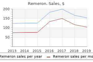
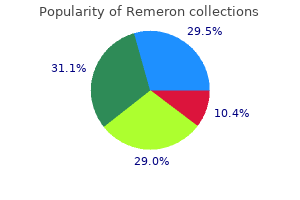
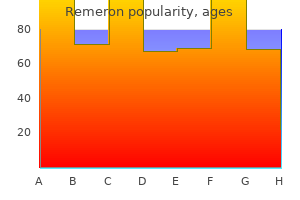
Cheap remeron online american express. 30 words you must AVOID in IELTS Writing.
Fixed superior humeral migration within the setting of a giant rotator cuff tear will lead to medications descriptions remeron 15mg lowest price inferior capsular contracture silent treatment purchase generic remeron on line. In these sufferers medicine lake california purchase remeron 15 mg on line, perform preoperative manipulation underneath anesthesia in ahead elevation to enhance the subacromial area obtainable symptoms 10 days post ovulation buy cheap remeron 15 mg on-line, thus facilitating the repair. Positioning Beach-chair position benefits that is an anatomic position that allows better orientation and understanding of shoulder anatomy whereas performing the repair. Examination under anesthesia is facilitated by stabilizing the scapula within the beach-chair position in contrast with the lateral position. The arm can be easily manipulated in surgery without the need to unhook it from a traction unit. Lateral decubitus place benefits Many surgeons believe that the lateral position improves visualization and maneuverability of the scope because of traction. It considerably improves inferior access to the glenohumeral joint, which makes it more easy to perform glenohumeral procedures but has little influence on subacromial procedures. Transient and permanent nerve damage has been reported due to traction within the lateral place. Consequently, we choose to perform all subacromial procedures, together with rotator cuff restore, in the beach-chair position. While the risks of surgical administration are well known, the risks of nonoperative remedy is in all probability not so obvious. Tear progression, muscle fatty infiltration and atrophy, and arthritis are potential irreversible dangers of nonoperative therapy of rotator cuff tears. Early surgical repair must be thought-about in all acute tears and any continual small or medium-sized tears in sufferers younger than age sixty five. These sufferers are at significant threat for growing the irreversible adjustments previously mentioned with prolonged nonoperative remedy. Consequently, the benefits of early surgical remedy mixed with the inherent risks of prolonged nonoperative therapy information us to early surgical repair. Preoperative Planning Tear measurement and chronicity will determine the problem of the restore, so careful preoperative imaging analysis is essential in surgical preparation. Our most popular starting posterior portal is barely more lateral than a normal posterior portal. This is completed to achieve higher visualization of the lateral larger tuberosity during repair. Also, a barely inferior place is preferred, since portals will migrate superiorly with shoulder swelling. Portals ought to be positioned low enough so that cannulas are launched parallel to the rotator cuff tendon. The portal should be placed at about the midpoint of the tear in small or medium-sized tears. A second lateral portal could be positioned in bigger tears with cannulas separated by a number of centimeters. Again, maintaining low portal placement is important so devices will be handed parallel to the tendon, allowing the greatest excursion of devices in the subacromial house. The anterolateral portal is mainly used as an adjunct portal for suture retrieval and storage. Repair Site Preparation A gentle tissue ablation gadget is used via the lateral portal to clear all the gentle tissue on the undersurface of the acromion extending posteriorly, together with the gentle tissue and fat around the scapular backbone. Soft tissue is removed from the higher tuberosity with a shaver, exposing cortical bone. Mobility of the torn tendon is assessed with a tissue grasper through the lateral portal. Anchor and Suture Placement Once the tear has been determined to be repairable, a medial row of suture anchors (5. For small and medium-sized tears, we routinely place two medial anchors on the degree of the anatomic neck.